Pathogenesis
Pathophysiology
Ulcerative Colitis
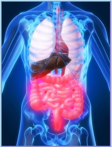
- It begins in the rectum (proctitis) and spreads proximally along the entire colon (pancolitis) in a continuous fashion.
- The mucosa of the rectum and the colon is hyperemic and edematous in the affected area.
- Multiple abscesses develop in the crypts of Lieberkuhn (intestinal glands).
- As the disease advances, the abscesses break through the crypts into the submucosa, leaving ulcerations.
- These ulcerations destroy the mucosal epithelium, causing bleeding and diarrhea – the loss of fluid and electrolytes caused by the decreased mucosal surface are for absorption.
- Protein loss through the stool is also evidenced due to breakdown of cells.
- Areas of inflamed mucosa can form pseudopolyps, tonguelike projections into the bowel lumen.
- Granulation tissue develops, and the mucosa musculature becomes thickened, shortening the colon.
Crohn’s Disease
-
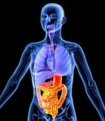 It is characterized by inflammation of segments of the GI tract.
- It can affect any part of the GI tract
- Most often seen in the terminal ileum and the colon.
- Involvement of the esophagus, the stomach, or the duodenum is uncommon.
- T-helper cell cytokines such as interleukin-12 and tumor necrosis factor (TNF) stimulate the inflammatory response, which begins in the intestinal submucosa and extends to all the layers of the bowel wall.
- Activated neutrophils and macrophages promote inflammation and cause tissue injury.
- The inflammation can affect some segments of the intestine but not others resulting in discontinuous skip lesions.
- Ulcerations are deep and longitudinal and penetrate between islands of inflamed edematous mucosa, causing the classic cobblestone appearance.
- Thickening of the bowel wall occurs, as well as narrowing of the lumen with stricture development – promotes obstruction.
- Abscesses or fistula tracts that communicate with other loops of bowel, the skin, the bladder, the rectum or the vagina may develop.
- Histologically, granulomas are present in 50% of clients and may be located in any layer of the bowel wall – giving the affected area a cobblestone appearance.
Previous Section: Pathogenesis
Home
Next Section:Clinical Manifestations
No comments:
Post a Comment